3d brain mri classification
Accurate and fully automatic brain tumor grading from volumetric 3D magnetic resonance imaging MRI is an essential procedure in the field of medical imaging analysis for full assistance of neuroradiology during clinical diagnosis. A very few features can be PDF BRAIN TUMOR CLASSIFICATION IN 3D-MRI USING FEATURES FROM RADIOMICS AND 3D-CNN COMBINED WITH KNN CLASSIFIER IAEME Publication - Academiaedu.
Diagnostics Free Full Text Mr Imaging Of Pediatric Brain Tumors Html
No packages published.

. Their 3D models are called 3D-VGG and 3D-ResNet and are widely used for 3D medical image classification study. Residual and Plain Convolutional Neural Networks for 3D Brain MRI Classification. In the first step a set of brain MRI scans is used to train the 3D-CNN.
I have used the IXI Brain MRI dataset that each image has 150 slices and it is available hereI downloaded the T1 images and used 80 percent of them for training and 20 for testvalidation. To examine the feasibility of applying CNN to classification of schizophrenia and controls based on structural Magnetic Resonance Imaging MRI we built 3D CNN models with different architectures and. Later I design a simple training workflow building on several well-established fr.
The knowledgeable regions are those areas of the brain which the 3D-CNN has mostly used to extract. Sergey Korolev Amir Safiullin Mikhail Belyaev Yulia Dodonova. Residual and plain convolutional neural networks for 3D brain MRI classification.
45 forks Releases No releases published. Convolutional Neural Networks for Classification of Alzheimers Disease and Mild Cognitive Impairment from 3D Brain MRI Images. Proper treatment planning and accurate diagnostics should be implemented to improve the life expectancy of the patients.
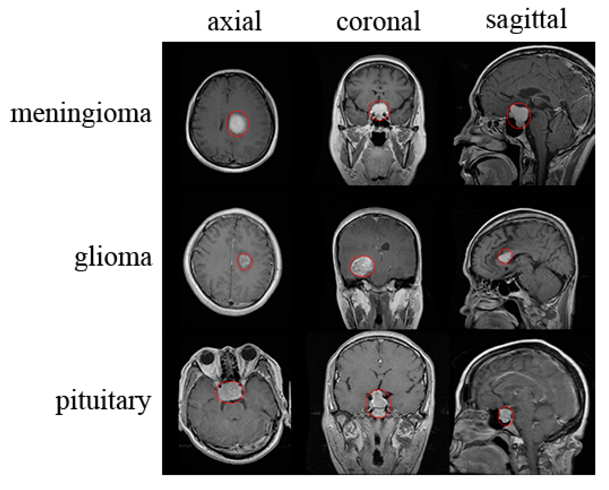
The framework of brain tumor segmentation is shown in Fig. Benign Tumor Malignant Tumor Pituitary Tumor etc. Mengjiao Hu Kang Sim.
Brain Tumors are classified as. CNN for MRI Gliomas Brain Tumor Classification J Digit Imaging. In the recent years there have been a number of studies that applied deep learning algorithms to neuroimaging data.
RSNA-MICCAI Brain Tumor Radiogenomic Classification which aims at brain tumor detection from 3D MRI scansI briefly describe the competition and provide data. Propose two 3D CNN architectures based on VGGNet and ResNet which is the first study to prove the manual feature extraction step for Brain MRI image classification is unnecessary. Code for Residual and Plain Convolutional Neural Networks for 3D Brain MRI Classification paper neuroml.
The proposed GABM method consists of two main steps. Korolev et al. Residual and Plain Convolutional Neural Networks for 3D Brain MRI Classification.
We use a UNet-like architecture for brain tumor segmentation and then apply a regular 3D CNN architecture for the tumor classification task. In the recent years there have been a number of studies that applied deep learning algorithms to neuroimaging data. Convolutional Neural Network CNN has been successfully applied on classification of both natural images and medical images but not yet been applied to differentiating patients with schizophrenia from healthy controls.
The input brain tumors are categorized into one of three sub-types. But classification becomes very difficult for the physician due to the complex structure of the brain. Brain MRI-based 3D Convolutional Neural Networks for Classification of Schizophrenia and Controls.
In the second step a genetic algorithm GA is applied to discover knowledgeable brain regions in the MRI scans. This post walks through our submission to the recent Kaggle competition. Given the subtle mixed and sparsely distributed brain atrophy patterns of schizophrenia the capability of automatic feature.
Ad Browse Discover Thousands of Medicine Book Titles for Less. Glioblastoma G oligodendroglioma O and astrocytoma A. Pipelines used in those studies mostly require multiple processing steps for.
Brain MRI Classification of AD Diagnosis Using Deep Learning. Proceedings of the 2017 IEEE 14th International Symposium on Biomedical Imaging ISBI 2017. Pipelines used in those studies mostly require multiple processing steps for feature extraction although modern advancements in deep learning for.

Evaluating Feasibility Of High Resolution T1 Perfusion Mri With Whole Brain Coverage Using Compressed Sense Application To Glioma Grading European Journal Of Radiology

Volbrain Automated Mri Brain Volumetry System

Glioblastoma Idh Wild Type Brain Mri In Adc Map A T2 Flair B And Download Scientific Diagram
Diagnostics Free Full Text Brain Tumor Detection And Classification On Mr Images By A Deep Wavelet Auto Encoder Model Html
Different Types Of Artifacts In Brain Mri A Chemical Shift B Download Scientific Diagram

Structural Brain Abnormalities In Adults With Congenital Heart Disease Prevalence And Association With Estimated Intelligence Quotient International Journal Of Cardiology
Sensors Free Full Text Mri Based Brain Tumor Classification Using Ensemble Of Deep Features And Machine Learning Classifiers Html

Standardized Brain Mri Protocol To Evaluate Patients In Whom Multiple Download Scientific Diagram
Mri Shows Brain Differences Among Adhd Patients Imaging Technology News
Brain Lesion Detection In Mri Images With Graph Cut Algorithms
Classification Of Brain Tumours In Mr Images Using Deep Spatiospatial Models Scientific Reports

Left Is Three Types Of Brain Tumor Mri Images T1 With Contrast T2 And Download Scientific Diagram

Brain Mri Obtained From A Sagittal Plane B Axial Plane And C Download Scientific Diagram

Brain Mri Scan Showing The Results Of The Parietal Vertex Craniotomy Download Scientific Diagram

7 T Magnetic Resonance Imaging In The Management Of Brain Tumors Magnetic Resonance Imaging Clinics

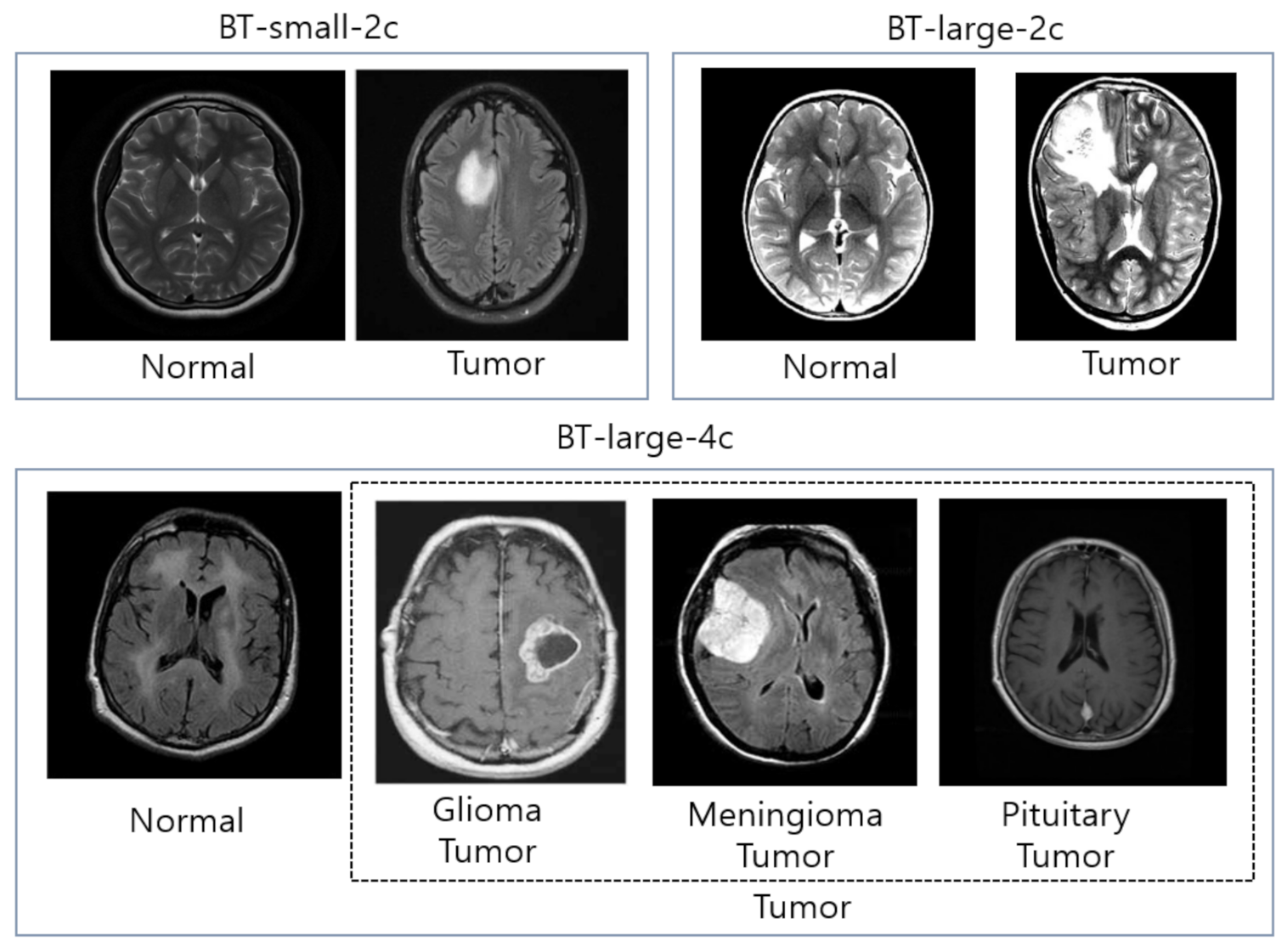
Sample Datasets Of Brain Tumor Mri Images Normal Brain Mri 1 To 4 Download Scientific Diagram

Incidental Findings On Brain Mri Arrows Indicate The Abnormalities In Download Scientific Diagram
Applied Sciences Free Full Text Classification Of Brain Tumors From Mri Images Using A Convolutional Neural Network Html

Brain Mri Obtained From A Sagittal Plane B Axial Plane And C Download Scientific Diagram